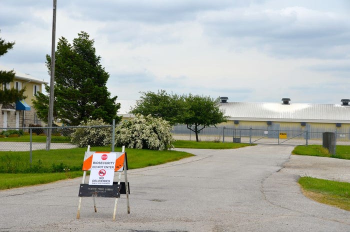
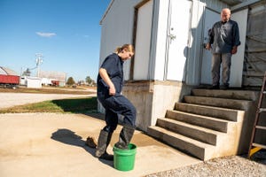
Biosecurity
thumbnail
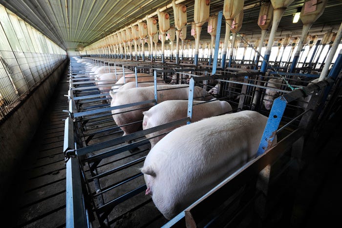
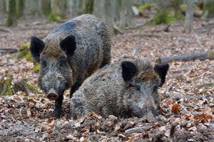
Livestock ManagementSqueal on Pigs secures additional $2.6 million in fundingSqueal on Pigs secures additional $2.6 million in funding
Proactive measures keep land, livestock safe; protect province’s pork industry.

.jpg?width=700&auto=webp&quality=80&disable=upscale)


.jpg?width=300&auto=webp&quality=80&disable=upscale)
Recent Headlines


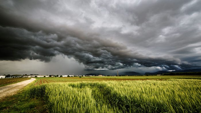
Enter a zip code to see the weather conditions for a different location.

Sep 27, 2023
Sep 27, 2023
Global Hog Industry Virtual Conference
Subscribe to Our Newsletters
National Hog Farmer is the source for hog production, management and market news