The secret life of PRRS virus … and what it means for PRRS diagnostics
Whether your goal is PRRS prevention, control or elimination, you need to use and interpret diagnostic test results in the context of the dynamics of PRRSV infection.
July 2, 2019

By Alexandra Henao-Diaz, David Baum, Luis Giménez-Lirola and Jeff Zimmerman, Iowa State University
If you are committed to raising healthy pigs, you are concerned about porcine reproductive and respiratory syndrome and have already implemented a strategy for preventing, controlling or eliminating PRRS virus in the pigs for which you are responsible. Whatever your chosen PRRS strategy, you need some method to track the success of your plan … or to detect failure. It should be clear by now that the majority of PRRS virus infections are subclinical, with clinical outbreaks of varying size interspersed over time in infected populations. If that were not true, we could simply look at pigs and know their PRRS virus infection status.
Therefore, using clinical signs to track the progress of a PRRS control plan is like using the dashboard warning light (a.k.a. “idiot light”) to monitor engine oil levels — good information, but a bit late. Even more problematic from the perspective of control and elimination, we now know that PRRS virus can circulate subclinically at low prevalence in sow herds for months longer that we had previously thought possible. In these sow herds, “leakage” of PRRS virus into piglets at farrowing results in infection of downstream production flows and puts PRRSV control or elimination goals at risk.
Since clinical signs are not reliable indicators of infection, the only option for monitoring the progress of PRRS control protocols and/or tracking the virus on production sites is routine testing. Whether or not you are currently testing, the goal of this article is to provide some insight into the biology of PRRS virus with the goal of making your testing plan more effective, less expensive, or both.
As described by Lopez and Osorio (2004), infection results in the presence of PRRS virus in tissues and blood (viremia) — the latter allows us to detect infection by testing blood samples for PRRS virus by RT-PCR. Over time, as the immune system responds and antibodies appear, virus is cleared from the circulatory system but remains in lymphoid tissues for a prolonged period of time. At this point in the infection, blood samples will test negative by PRRS reverse transcription polymerase chain reaction, even though animals still carry infectious virus and can infect other pigs. Carrier pigs are the curse of elimination projects because of their role in the reappearance of PRRS virus in presumed-negative populations. Eventually, the virus is cleared from all tissues and the pigs become truly PRRS virus negative, although we do not currently have the means to determine exactly when that happens.
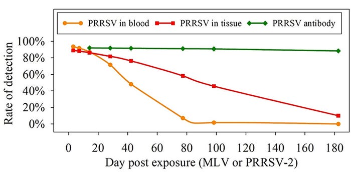
This diabolically clever strategy optimizes the probability that the virus will continue to circulate in pigs and confound our best efforts to control it, but what exactly does it mean for PRRSV diagnostics? To answer this question, approximately 3,800 individual diagnostic test results were compiled from 19 refereed publications describing PRRSV infection in pigs. These data were used to estimate the rates of viremic pigs, carrier pigs and antibody-positive pigs over time post exposure.
As Figure 1 shows, the probability of detecting PRRS virus infection changes over time as a function of specimen and diagnostic target. For example, at 98 days post exposure the analysis predicted that fewer than 2% would be serum RT-PCR positive, nearly 50% of pigs would carry infectious virus in tissues, and more than 90% would be enzyme-linked immunosorbent assay antibody positive. These results have important diagnostic implications. In particular, Figure 1 shows that sole reliance on serum RT-PCR testing must necessarily produce increasing numbers of false negative results over time. Likewise, it shows that RT-PCR negative results from antibody positive pigs is not a diagnostic contradiction. To the contrary, the expected diagnostic profile of a PRRS virus carrier would be ELISA positive and RT-PCR negative, not because the tests are deficient but because of the dynamics of the infection.

Whether your goal is PRRS prevention, control or elimination, you need to use and interpret diagnostic test results in the context of the dynamics of PRRSV infection. No single testing plan will meet the objective of every situation, but the results of this analysis show that the best overall detection strategy will coordinate the use of RT-PCR to detect acute PRRSV infections and ELISA thereafter (Figure 1).
References
Lopez OJ, Osorio FA. 2004. Role of neutralizing antibodies in PRRSV protective immunity. Vet Immunol Immunopathol 102:155-163.
Sources: Alexandra Henao-Diaz, David Baum, Luis Giménez-Lirola and Jeff Zimmerman, who are solely responsible for the information provided, and wholly own the information. Informa Business Media and all its subsidiaries are not responsible for any of the content contained in this information asset.
You May Also Like


