Subscribe to Our Newsletters
National Hog Farmer is the source for hog production, management and market news
Identifying the culprit in the line-up of suspects requires more than casual observation. A diagnostic evaluation by a veterinarian or pathologist is more likely to ferret out the correct answer, much like accumulating evidence in a mystery novel, says Kent Schwartz, DVM, diagnostician at the Iowa State University Veterinary Diagnostic Laboratory

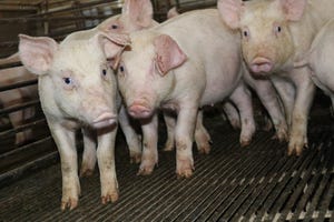
Ileitis is a swine intestinal disease that costs pork producers in pig deaths, treatments, preventatives and perceptions regarding welfare and antimicrobial use. More subtly, it costs in lost feed efficiency and growth rates that are often not apparent until the damage is done and profit margins are diminished.
Identifying the culprit in the line-up of suspects requires more than casual observation. A diagnostic evaluation by a veterinarian or pathologist is more likely to ferret out the correct answer, much like accumulating evidence in a mystery novel, says Kent Schwartz, DVM, diagnostician at the Iowa State University Veterinary Diagnostic Laboratory (ISU-VDL).
A wide range of infectious agents, mostly bacterial, were identified as enteric agents in pigs older than 6 weeks of age at the ISU-VDL in 2010. In Figure 1, ileitis caused by Lawsonia intracellularis ranks second in relative frequency of diagnosis, while salmonella enteritis ranks highest. Together, these two bacteria account for nearly 40% of all cases, Schwartz said during a talk at the World Pork Expo in June. Noteworthy is the increasing frequency of diagnosis of Brachyspira (similar to swine dysentery and often called spirochaetal colitis) as a cause of grow-finish diarrhea.
Many of the enteric pathogens are endemic, meaning they are common infections and can cause disease when the conditions are right. While the “tip of the iceberg” cost of deaths and medication can range from $3-
11/head, the “invisible part of the iceberg” can be more costly: an additional $2-11/head due to subclinical losses of feed conversion, gain and weight variation or carcass quality at marketing, he notes.
Schwartz says there doesn’t seem to be much difference in seasonality for salmonella, based on data for 2003-2010, but over a third of ileitis cases were identified during the July-September period, he notes. Salmonella choleraesuis used to be the most feared salmonella infection of pigs, but in the last decade, Salmonella typhimurium far outranks all other salmonella serotypes in frequency as a cause of disease.
Pigs affected with ileitis, salmonella or Brachyspira can all look quite similar. Salmonella and ileitis are common infections in most herds, but they are not frequently found as simultaneous causes of disease in individual pigs. Each is an important cause of production losses and disease, and it only takes one of them to cause performance losses in pigs. Because these agents have similar clinical signs and effects, laboratory testing is frequently sought to confirm the actual cause of disease.
Data from ISU-VDL shows age trends and number of cases for salmonella and ileitis. Salmonella is a more common disease in the nursery phase and declines to lower levels in the finisher, while cases of ileitis reflect a fairly opposite trend.
Accurate, timely and useful disease diagnosis begins with close observations at the farm and individual pig level, Schwartz stresses. After getting an accurate diagnosis by a laboratory submission, production data should be evaluated for herd impact and to monitor the efficacy of interventions.
Remember that salmonella, ileitis, Brachyspira and a host of other enteric pathogens can strike at any age postweaning, he points out.
Ileitis Diagnosis, Prevention
In the United States, prevalence of ileitis on hog farms remains at 96%, based on serum antibody samples. However, the prevalence varies between farms and production flows.
Clinical forms of ileitis include an acute form (diarrhea, debilitation, deaths) and a more chronic form (milder diarrhea, slow growth).
With subclinical disease, diarrhea may not be seen; there are more subtle effects of infection, including compromised feed efficiency and growth rate. Accurate diagnosis of clinical disease often requires laboratory testing. Diagnosis of subclinical effects definitely requires laboratory testing, coupled with scrutiny of production records.
With clinical disease, diarrhea is the first sign, and gross lesions may suggest ileitis, but histopathology (microscopic examination) with immunohistochemistry staining (use of antibody to detect organisms with a microscope) is considered the gold standard to confirm that the organism is causing disease, Schwartz said during a World Pork Expo press conference sponsored by Boehringer Ingelheim Vetmedica (BIVI) Inc. Testing feces by polymerase chain reaction (PCR) can find the organism in infected pigs, but it doesn’t confirm that disease is present, he says.
Vaccination for ileitis (Enterisol Ileitis by BIVI) is approved for piglets at 3 weeks of age or older. To be effective, vaccine must be given orally at least three weeks prior to the time pigs are expected to become infected. Since clinical signs of disease occur 2-3 weeks after infection, a six-week window between vaccination and expected clinical signs is necessary for good vaccine efficacy. Infection often occurs around 10-16 weeks of age, so the “default” timing of vaccination is usually mid- to late-nursery stage. If pigs are properly vaccinated, the duration of immunity carries to at least 26 weeks of age, Schwartz says.
Subclinical infections can be detected by testing feces with PCR or by the use of serological (blood) tests. These methods can confirm if infection is occurring and, if performed at different ages, can determine when the infection occurs — useful information when planning intervention strategies. Usually, this involves sampling 15-30 pigs at various ages.
For example, if serology suggests more than 10% of pigs are positive at 12 weeks of age, then vaccination should be placed no later than 6 weeks of age (keeping in mind the 5-6 week window between infection and seroconversion).
It is important to select the right ages and types of animals for laboratory testing. Your veterinarian can help determine the best type of testing strategy for your herd.
You May Also Like
Enter a zip code to see the weather conditions for a different location.