Subscribe to Our Newsletters
National Hog Farmer is the source for hog production, management and market news
Mhp transmits slowly within exposed populations. Even during outbreak events, the proportion of positive gilts at one time never exceeded 52%.
June 4, 2018

By M.J Clavijo, Pig Improvement Co. and Iowa State University College of Veterinary Medicine Department of Veterinary Diagnostic and Production Animal Medicine; C Johnson, Carthage Veterinary Services; A. Farkas, Carthage Veterinary Services; and J.P. Cano, Iowa State University College of Veterinary Medicine Department of Veterinary Diagnostic and Production Animal Medicine
Jim Lowe adequately described the main challenge with Mycoplasma hyopneumoniae in the field1. Gilts, regardless of their infection status (positive or negative) are likely the main cause of breeding herd health destabilization and the source of infection for piglets. Proper gilt acclimation hinges upon early Mhp exposure (natural or intentional). This will allow sufficient time to minimize bacterial shedding and build up immunity, successfully protecting their piglets and the health of the breeding herd.
As such, a key factor here is the duration of shedding of the population, since it will determine the appropriate timing of introduction of gilts into the herd and the duration of herd closure in elimination efforts. Though the duration of shedding in experimentally infected pigs has been assessed, the epidemiology of Mhp in naturally infected gilt populations remains largely unknown.2 The objective of this study was to describe the pattern of Mhp infection and persistence in a naturally exposed gilt population.
The study
The farm enrolled in this study had experienced an Mhp outbreak in August 2016, after being negative for more than five years. In this prospective cohort study, a group of 63 unvaccinated gilts were randomly selected at 21 days of age, tagged and sampled via laryngeal or tracheal sampling weekly for five weeks and monthly afterwards. Serum samples were collected at 21, 110 and 140 days of age. A final sampling occurred during the farrowing stage, where a tracheal sample was collected from study sows and five of their piglets. Tracheal samples were collected by inserting the end of a post-cervical artificial insemination rod into the trachea. All laryngeal and tracheal swabs were tested by quantitative Mhp polymerase chain reaction and serum samples were tested for Mhp antibodies by IDEXX ELISA.
Despite the recent Mhp outbreak, the prevalence at weaning was low (11%). While such slow transmission is a common feature within the species, it is hypothesized that the lower prevalence could be due to the administration of in-feed antibiotic in the sows, a less virulent Mhp strain or the lack of sensitivity of the laryngeal method for early detection. The prevalence at 80, 110, 140 and 170 days of age was 15%, 30%, 29% and 4%, respectively. The early decline in detection at 170 days led to the decision to implement a more invasive and sensitive diagnostic method.4 The detection of Mhp in laryngeal swabs was 9% compared to 42% in tracheal samples at 200 days of age, and 5% compared to 52% using tracheal samples at 215 days. The latter sampling method appears to be a more sensitive methodology for detection of Mhp during chronic infection.3,4 The prevalence decreased from 33% at 245 days to 6% at 305 and 0% at 325 days. In this study, at least one gilt was PCR positive 284 days after initial detection, however most gilts were detected positive for less than 200 days, suggesting a potential for shorter herd closure periods and thus more cost-effective elimination programs. At farrowing, 20 study sows and five of their due-to-wean piglets tested PCR negative. This information suggests that gilts cleared infection within normal Mhp elimination timelines without intentional exposure.

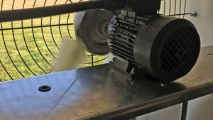
Tracheal samples of pigs were collected by inserting the end of a post-cervical artificial insemination rod into the trachea.
Diagnostic tools are not perfect
A total four gilts (6.3%) remained PCR negative throughout the study; however; there was evidence of seroconversion in 100% of the gilts by 140 days. This reveals that although 93% of gilts in this study showed at least one PCR positive laryngeal or tracheal sample, not all exposed gilts were detected positive, even after consecutive samplings. The seroprevalence at weaning was 100%, evidencing a consistent transfer of maternal antibodies from sow to piglet. The seroprevalence at 110 days was 60%. From these seropositive gilts, 39% were PCR negative using laryngeal swabs. Seroconversion is usually seen between four and eight weeks after infection, consequently these results would indicate that the laryngeal swabbing method was not effective for detection of infection in previous samplings. In contrast, 40% of the gilts were serologically negative at 110 days of age, and from these, 27% had previously tested PCR positive by laryngeal swabs between 21 and 49 days of age.
This shows the variability in seroconversion for Mhp and supports the fact that antibody response is a lagging indicator for exposure. The seroprevalence at 140 days revealed that 100% of the gilts had seroconverted, still 30% of seropositive gilts were not detected PCR positive by laryngeal swabs after eight different sampling events. The lack of sensitivity of the sampling methods should be taken into account when designing surveillance programs for Mhp (i.e. determining day “zero” or detection prior to reopening a herd).
Take-home messages
1. Shedding and exposure of piglets to Mhp in the farrowing room, even under recent introduction into a negative herd, can be low.
2. Mhp transmits slowly within exposed populations. Even during outbreak events, the proportion of positive gilts at one time never exceeded 52%.
3. The peak of acute infection detected by laryngeal swabs was observed at 49 days of age (39%). The peak of chronic infection was observed at 215 days using tracheal swabs (52%).
4. Shorter duration of detection (less than 200 days) suggests potential for shorter herd closure periods and potential for more cost-effective elimination programs.
5. Laryngeal swabs appear to lack sensitivity for Mhp detection in mid- to late-infection, as evidenced by seroconversion coupled with negative laryngeal swabs by PCR. Hence, tracheal samples appear to be more sensitive diagnostic tool compared to laryngeal swabs.
6. Mhp antibody response is a lagging indicator for exposure.
7. Early natural exposure of gilts can lead to successful Mhp acclimation.
References
You May Also Like
Enter a zip code to see the weather conditions for a different location.