UVC can drastically reduce viable PRRSV and PEDV within a 5 minute exposure time.
October 31, 2017

Sponsored Content
Reduction in Swine Pathogen Numbers by a UVC germicidal chamber
Porcine reproductive and respiratory syndrome virus PRRSV is recognized as a major challenge to the pork industry, imposing an estimated $560 million in losses annually (Neumann et al., 2005, Rossow, 1998). Another virus, porcine epidemic diarrhea virus PEDV, has more recently resulted in one of the most devastating outbreaks the pork industry had endured (Lee, 2015). These viruses, like many other pathogens, may be transmitted by the import of contaminated objects (fomites) into biosecure facilities (Dee et al., 2002, 2004, Otake et al., 2002). Therefore disinfection of fomites represents a key biosecurity measure in any swine operation.
Germicidal effects of ultraviolet UV irradiation were discovered more than a century ago. Short-wavelength ultraviolet C UVC light has been determined the most effective. The entire UVC range of ultraviolet radiation is germicidal and falls into a subset of light called ultraviolet germicidal irradiation UVGI. UVGI disrupts (or mutates) the DNA and RNA of bacteria, viruses and a majority of fungal pathogens — destroying their ability to multiply and cause disease. In this study, we evaluated the first UVC germicidal pass-through chamber designed for the agricultural market, Once Inc.’s BioShift UV chamber. The BioShift exhibited large reductions in PEDV as well as PRRSV. Suggested considerations for use are discussed.
Methods
BioShift construction:
A commercial heavy-duty pass-through chamber was constructed. Harsh atmospheric conditions, usage cycles and human interaction in agricultural buildings/barns were considered as additional factors for choosing the chamber’s materials. There are two sides to the chamber, a dirty (street), and a clean (biosecure) side. Safety switches were installed on both sides to ensure powering off the germicidal lamp light source when one or both doors would be opened. All exposed electrical and mechanical parts to UV radiation are made from UVC-resistant materials. 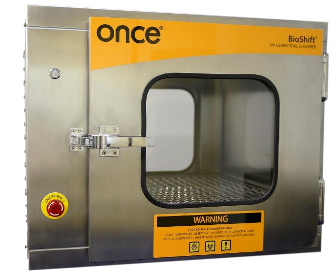
All exposedelectrical and mechanical parts to UV radiation are made from UVC-resistant materials. Image of the BioShift Cabinet.
Virus viability assays
PEDV virus strain Colorado 2013 isolate, was obtained from the National Veterinary Services Laboratories NVSL, Ames, Iowa. PRRS virus strain NVSL, was obtained from the University of Kentucky. An organic soil load of 5% fetal bovine serum FBS was used throughout the experiments.
For each exposure time and exposure condition, duplicate carriers (100 X 15 mm sterile glass petri dishes) were inoculated with a 200 μL aliquot of virus suspension and allowed to air dry. The exposure times were 1 minute, 2 minutes and 5 minutes and the exposure conditions were direct and indirect/shadow exposure to UVC light. At the end of each exposure time, a 2.00 mL aliquot of test medium was added to each carrier and they were individually scraped with a cell scraper to resuspend the contents (101 dilution). The contents of each carrier were transferred to individual snap cap tubes and assayed for infectivity and/or cytotoxicity. Appropriate virus, cytotoxicity and neutralization controls were run concurrently.
Results
BioShift specifications
A pass-through chamber was constructed using germicidal lamps (Figure 1). UVC intensities were measured at 16 points within the BioShift®, and were used to generate a heat map (Figure 2). Intensity was highest near the bulbs and diminished as the distance from the bulb increased. On average, the UV irradiance levels were 180-280 μW/cm2 along the tray surface.
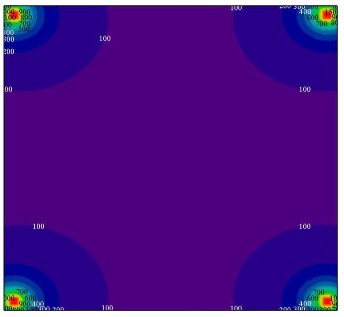
UVC light distribution within the BioShift The image is a cross-section, viewed as if the door were open. Intensity units are in μW/cm2.
Determination of input virus titers.
The PRRS and PED virus stocks were diluted 10-fold and assayed for infectivity and/or cytotoxicity in their respective host cell lines. Based on these control results, the 50% tissue culture infective dose (TCID50)/100 μL for the PRRSV input was determined to be 106.00. The TCID50/200 μL for the PEDV input was determined to be 105.50.
Determination of viral killing rates
Samples were exposed to UVC in the BioShift or one, two, or five minutes, and were compared to untreated control samples whereby the BioShift was left off for the same amount of time. UVC light was either in direct contact with the sample, or was shadowed by a petri dish lid. Viral numbers were quantified according to the TCID50.
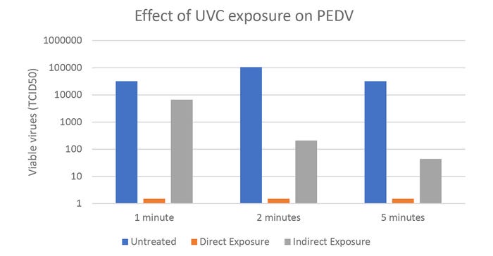
Quantification of viable PEDV treated with direct or indirect UVC radiation for one, two and five minutes in the BioShift Log10 reductions for direct exposures were ≥4.00, ≥4.52, and ≥4.00 for one, two, and five minutes, respectively. Log10 reductions for indirect exposures were 0.68, 2.70, and 2.86. Note that the vertical axis is on a logarithmic scale.
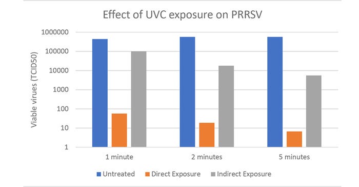
Quantification of viable PRRSV treated with direct or indirect UVC radiation for one, two and five minutes in the BioShift. Log10 reductions for direct exposures were 3.89, 4.48, and ≥4.93 for one, two and five minutes respectively. Log10 reductions for indirect exposures were 0.64, 1.50, and 2.01. Note that the vertical axis is on a logarithmic scale.
This study demonstrated that UVC drastically reduced viable PRRSV and PEDV within a five minute exposure. Since direct exposure was more effective than indirect, objects should be spread out to maximize the amount of UVC light hitting the object. As light intensity follows the inverse squares law, the objects closer to the UV bulbs will result in higher disinfection rates. According to the time course analysis, the majority of killing occurred within the first two minutes of exposure. Weighing the benefits of additional exposure time with the drawback of requiring the user to wait additional time, we recommend five minutes exposure for most applications.
The dose of UVC given off by five minutes of exposure in the BioShift is harmless to most materials. Over repeated exposure, some plastics may appear yellow and may become brittle. Maximum germicidal activity will be achieved for objects with hard surfaces rather than soft, porous surfaces.
This study serves as a proof of concept looking at two common swine viruses. As evidenced by the differences in killing between PRRSV and PEDV, some pathogens could be more susceptible to UV radiation than others. Differing susceptibilities to UVC can be attributed to differences in DNA repair enzymes. For example, Arrage, et al. 1993, showed that different bacteria showed different sensitivities to UVC. Most bacteria were reduced by 90% with a fluence of 3-4 mW*s*cm-2 UVC radiation. Five minutes exposure in the BioShift results in 54-84 mW*s*cm-2 UVC radiation.
Altogether this study demonstrates that UVC irradiation by the BioShift can serve as an effective biosecurity tool. Together with other biosecurity best practices, reducing the import of pathogens into biosecure facilities will result in fewer disease outbreaks.
For a video demonstration, click here.
References
Arrage, A.A., Phelps, T.J., Benoit, R.E., White, D.C., 1993. Survival of subsurface microorganisms exposed to UV radiation and hydrogen peroxide. Appl. Environ. Microbiol. 59, 3545–3550.
Bolton, J.R., Cotton, C., Christine Anne, 2011. The Ultraviolet Disinfection Handbook. American Water Works Association.
Cutler, T.D., Wang, C., Hoff, S.J., Kittawornrat, A., Zimmerman, J.J., 2011. Median infectious dose (ID50) of porcine reproductive and respiratory syndrome virus isolate MN-184 via aerosol exposure. Vet. Microbiol. 151, 229–237. doi:10.1016/j.vetmic.2011.03.003
Cutler, T.D., Wang, C., Hoff, S.J., Zimmerman, J.J., 2012. Effect of temperature and relative humidity on ultraviolet (UV 254) inactivation of airborne porcine respiratory and reproductive syndrome virus. Vet. Microbiol. 159, 47–52. doi:10.1016/j.vetmic.2012.03.044
Dee, S., Deen, J., Rossow, K., Wiese, C., Otake, S., Joo, H.S., Pijoan, C., 2002. Mechanical transmission of porcine reproductive and respiratory syndrome virus throughout a coordinated sequence of events during cold weather. Can J Vet Res 66, 232–239.
Dee, S.A., Deen, J., Otake, S., Pijoan, C., 2004. An experimental model to evaluate the role of transport vehicles as a source of transmission of porcine reproductive and respiratory syndrome virus to susceptible pigs. Can J Vet Res 68, 128–133.
Dee, S., Otake, S., Deen, J., 2011. An evaluation of ultraviolet light (UV254) as a means to inactivate porcine reproductive and respiratory syndrome virus on common farm surfaces and materials. Vet. Microbiol. 150, 96–99. doi:10.1016/j.vetmic.2011.01.014
Downes, A., Blunt, T.P., 1878. On the Influence of Light upon Protoplasm. Proc. R. Soc. Lond. 28, 199–212. doi:10.1098/rspl.1878.0109
Lee, C., 2015. Porcine epidemic diarrhea virus: An emerging and re-emerging epizootic swine virus. Virol J 12. doi:10.1186/s12985-015-0421-2
Neumann, E.J., Kliebenstein, J.B., Johnson, C.D., Mabry, J.W., Bush, E.J., Seitzinger, A.H., Green, A.L., Zimmerman, J.J., 2005. Assessment of the economic impact of porcine reproductive and respiratory syndrome on swine production in the United States. J. Am. Vet. Med. Assoc. 227, 385–392. doi:10.2460/javma.2005.227.385
Otake, S., Dee, S.A., Rossow, K.D., Deen, J., Han, S.J., Molitor, T.W., Pijoan, C., 2002. Transmission of porcine reproductive and respiratory syndrome virus by fomites (boots and coveralls). Journal of Swine Health and Production 10, 59–65.
Rossow, K.D., 1998. Porcine Reproductive and Respiratory Syndrome. Vet. Pathol. Online 35, 1–20. doi:10.1177/030098589803500101
Song, D., Park, B., 2012. Porcine epidemic diarrhoea virus: a comprehensive review of molecular epidemiology, diagnosis, and vaccines. Virus Genes 44, 167–175. doi:10.1007/s11262-012-0713-1
The Nobel Prize in Physiology or Medicine 1903 [WWW Document], n.d. URL http://www.nobelprize.org/nobel_prizes/medicine/laureates/1903/ (accessed 7.27.16).
About the Author(s)
You May Also Like

