Subscribe to Our Newsletters
National Hog Farmer is the source for hog production, management and market news
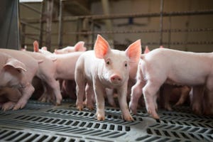
Weaning is a stressful event for the young pig. Weaning before three weeks results in long-term negative effects on immune system and mucosal development.
April 18, 2017

By Christopher Chase, South Dakota State University
Stressors such as early and abrupt weaning of the piglet, between 17 and 28 days of age, place enormous stress on the piglet’s gastrointestinal tract (gut), leading to perturbations in gut microbiota, host physiology and mucosal immune function. This often results in post-weaning diarrhea. It is characterized by diarrhea and growth reduction attributable to enterotoxigenic strains of E. coli. The health of the mucosa epithelium, the cells that line the gastrointestinal tract, is important not only for the growth and development of the pig through secretion and absorption but these epithelial cells (aka enterocytes) are the largest immune organ in the pig, and are the first responder to microorganisms in the gut.
The goblet cells that are part of the mucosa secrete mucous and mucins that provide the initial barrier (Figure 1)1. Mucosa epithelial cells also contribute mucins to this barrier2. This barrier also contains antimicrobial peptides produced by the enterocytes (Figure 1). These AMP are induced by the microbiota and metabolites like short chain fatty acids in the intestinal tract. AMP are also induced by the immune natural killer and LTi cells (Figure 1)2.
The AMP (aka host defense peptides) are an important family of molecules that helps to form a chemical barrier to limit infection at epithelial surfaces and attack invading bacteria. In pigs, more than 30 HDPs have been identified and characterized3. The AMPs are relatively small cationic peptides and are found predominantly at mucosal surfaces and in phagocytic cells. They vary in structure and anti-microbial activity and can be functionally differentiated during cellular processing. The concentration of some HDPs increases in response to inflammation or microbial infection. Secretory IgA is produced when dimeric IgA is secreted by the plasma cells in the lamina propria and is transported to the mucosal surface of the epithelial cell (Figure 1)2.
The commensal microbiota in the gut is important for the production of IgA3. Secretory IgA plays an important role in immunity at mucosal surfaces by causing infectious agents to stick together, preventing attachment of infectious agents to epithelial cells, and neutralizing toxins. The inner mucous layer along with the AMP and sIgA form a “killing zone” that few pathogens or commensals have evolved strategies to penetrate (Figure 1). This “killing zone,” along with the tight junctions that knit the enterocytes, forms a “barrier” against E. coli and other enteric pathogens.

Figure 1: Mucosal defenses. Distinct subpopulations of intestinal epithelial cells (IECs) are integrated into a continuous, single cell layer that is divided into apical and basolateral regions by tight junctions. Epithelial cells sense the microbiota to induce the production of antimicrobial peptides (AMPs). Goblet cells produce mucin, that is organized into a dense, more highly cross-linked inner proteoglycan gel that forms an adherent inner mucous layer, and a less densely cross-linked outer mucous layer. The outer layer is highly colonized by constituents of the microbiota. The inner mucous layer is largely impervious to bacterial colonization or penetration due to its high concentration of bactericidal AMPs, as well as commensals specific secretory IgA (sIgA) in the inner mucous layer to form the “killing zone”. Responding to the microbiota, innate lymphoid cells (ILC), lymphoid tissue inducer cells (LTi) and natural killer cells (NK), produce cytokines, which stimulate AMP production and maintain the epithelial barrier. A similar scheme is seen in respiratory mucosa. Adapted from Maynard et al., Nature 489, 231–241, 2012
Other components of the immune response may also be important in protection against various types of infection at mucosal surfaces. The immigration of neutrophils into the lumen of the gut and their subsequent destruction results in an increased concentration of lactoferrin, lysozyme and cationic proteins. These substances may also contribute to immunity to bacterial infections in the gut2.
Weaning is a stressful event for the young pig. Weaning before three weeks results in long-term negative effects on immune system and mucosal development. Early weaning at 17-21 days results in prolonged increase in the stressed hormone, corticotrophin-releasing factor and increases mast cells that produce histamine that results in diarrhea and leaky gut4.
The stress of being removed from the sow, age at weaning, mixing into a new environment and abruptly withdrawn from the sow’s milk results in the gut microbial ecosystem and lowered defenses against pathogen entry, lead to increased risk of disease, in particular, post-weaning diarrhea3. Post-weaning diarrhea is characterized by reductions in healthy commensal bacteria, i.e. Lactobacillus spp, and increases in pathogenic E. coli3. This leads to “dysbiosis” — when the good bacteria decrease and the harmful organisms like E. coli3,4 can take over. Dysbiosis is not just the loss of microbiota, it results in depletion of the “killing zone” (Figure 1) — the mucous layer becomes thinner and the amount of sIgA and AMP declines to allow the barrier to become weakened, allowing pathogenic enterotoxigenic E. coli to come in contact with mucosa and cause disease (Figure 2).

Figure 2: Gut microbiota and their products shape the development of epithelial cells and immunity. (A) Supplementing piglets with prebiotics or probiotics can increase microbial diversity, which can help to exclude pathogenic microbes. At weaning, feeding complex diets containing fermentable carbohydrates may further increase microbial diversity, helping to exclude pathogens from colonizing and causing disease. (B) Failure to supplement diets with prebiotics and probiotics or feeding simple diets at weaning can exacerbate colonization of pathogens, causing villus atrophy, inflammation, diarrhea, and growth reduction. Adapted from Fouhse et al. Animal Frontiers 6, 30–36, 2016.
One major factor leading to the dysbiosis and post-weaning diarrhea is low feed and water intake3. Post-weaning anorexia and dehydration lead to structural changes to the intestine, including villus atrophy and crypt hyperplasia, which further lead to gut barrier dysfunction4. Early weaning before 20 days of age results in an impaired innate immune response with lower numbers of neutrophils and inflammatory cytokines that are necessary to control post weaning E. coli infections4. To make things go from bad to worse, the reduction of lactic acid-producing bacteria (Lactobacillus) during weaning raises intestinal pH, increasing disease susceptibility because low gut pH is bactericidal to E. coli3. Applied research in weaning pigs using prebiotics, organic acids, probiotics and increased fiber improves gut health and piglet health (Figure 2)5,6,7.
These improvements occur through mechanism like increasing the kill zone, enhancing the thickness of the mucous, increasing levels of IgA and enhancing the anti-inflammatory response of the immune system. The other effects of the probiotics and prebiotics increase symbiotic microbes and decrease opportunistic pathogens, minimizing dysbiosis and “gut dysfunction.”
References
Pelaseyed, T., Bergström, J.H., Gustafsson, J.K., Ermund, A., Birchenough, G.M.H., Schütte, A., van der Post, S., Svensson, F., Rodríguez-Piñeiro, A.M., Nyström, E.E.L., Wising, C., Johansson, M.E.V., Hansson, G.C., 2014. The mucus and mucins of the goblet cells and enterocytes provide the first defense line of the gastrointestinal tract and interact with the immune system. Immunol. Rev. 260, 8–20. doi:10.1111/imr.12182
Maynard, C.L., Elson, C.O., Hatton, R.D., Weaver, C.T., 2012. Reciprocal interactions of the intestinal microbiota and immune system. Nature 489, 231–241. doi:10.1038/nature11551
Fouhse, J.M., Zijlstra, R.T., Willing, B.P., 2016. The role of gut microbiota in the health and disease of pigs. Animal Frontiers 6, 30–36. doi:10.2527/af.2016-0031
McLamb, B.L., Gibson, A.J., Overman, E.L., Stahl, C., Moeser, A.J., 2013. Early weaning stress in pigs impairs innate mucosal immune responses to enterotoxigenic E. coli challenge and exacerbates intestinal injury and clinical disease. PLoS ONE 8, e59838. doi:10.1371/journal.pone.0059838
Grilli, E., Tugnoli, B., Passey, J.L., Stahl, C.H., Piva, A., Moeser, A.J., 2015. Impact of dietary organic acids and botanicals on intestinal integrity and inflammation in weaned pigs. BMC Vet. Res. 11, 96. doi:10.1186/s12917-015-0410-0
Heinritz, S.N., Weiss, E., Eklund, M., Aumiller, T., Louis, S., Rings, A., Messner, S., Camarinha-Silva, A., Seifert, J., Bischoff, S.C., Mosenthin, R., 2016. Intestinal Microbiota and Microbial Metabolites Are Changed in a Pig Model Fed a High-Fat/Low-Fiber or a Low-Fat/High-Fiber Diet. PLoS ONE 11, e0154329. doi:10.1371/journal.pone.0154329
Vlasova, A.N., Kandasamy, S., Chattha, K.S., Rajashekara, G., Saif, L.J., 2016. Comparison of probiotic lactobacilli and bifidobacteria effects, immune responses and rotavirus vaccines and infection in different host species. Veterinary Immunology and Immunopathology 172, 72–84. doi:10.1016/j.vetimm.2016.01.003
You May Also Like
Enter a zip code to see the weather conditions for a different location.